Ophthalmic investigations

Eye examinations for diagnosis and monitoring

Diagnostic examinations complement the eye consultation with images and objective measurements. The ophthalmologist selects the tests appropriate to the clinical question and interprets the findings together with your symptoms, history and eye examination.

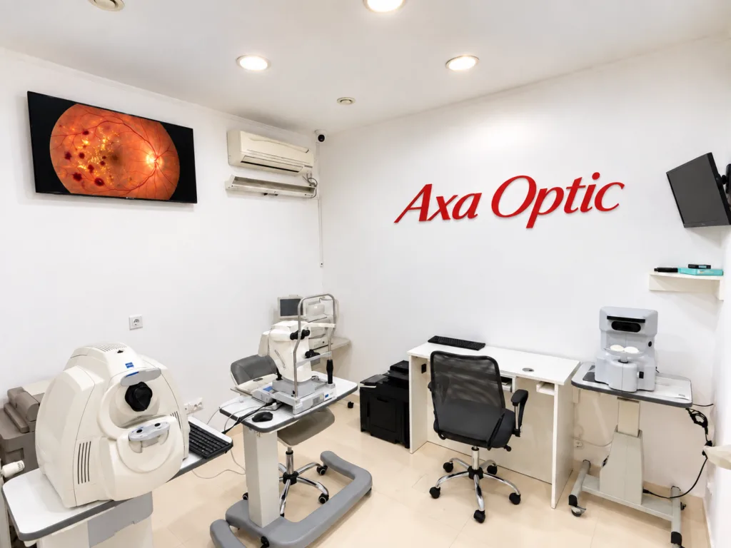
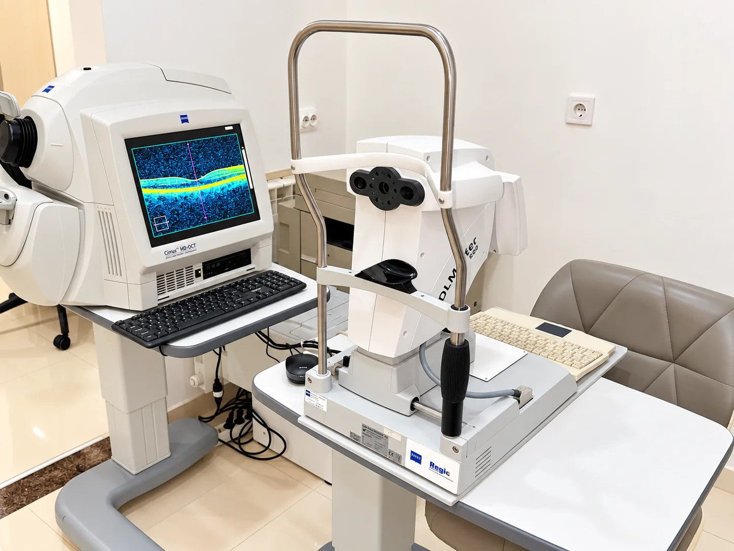
Axa Optic ophthalmic investigations room in Bârlad
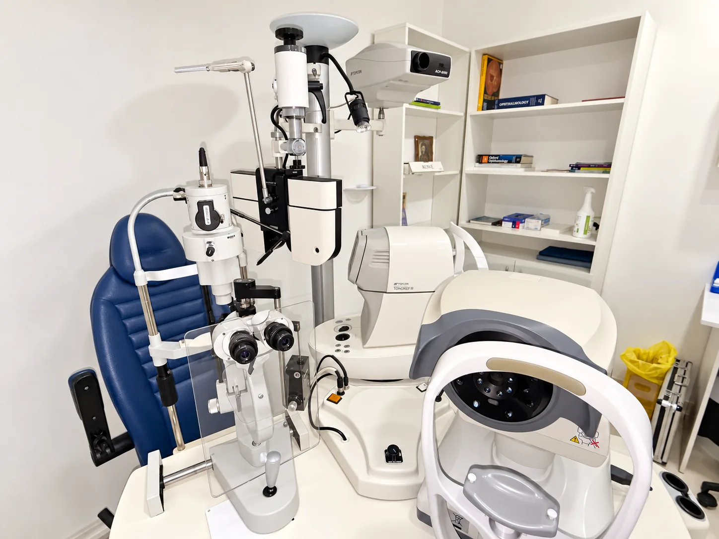
Diagnostic equipment at the Axa Optic ophthalmology clinic
What to expect
  • The examinations described here do not involve incisions or injections.
  • Examination time varies according to the test and patient cooperation.
  • Pupil dilation is sometimes necessary.
  • Results are interpreted by a physician in clinical context.
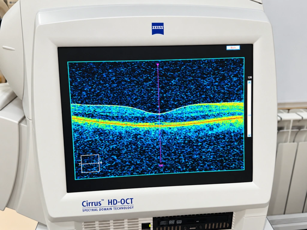
Axa Optic — Optical coherence tomography (OCT)
Optical coherence tomography (OCT)
Additional example: Optical coherence tomography (OCT)
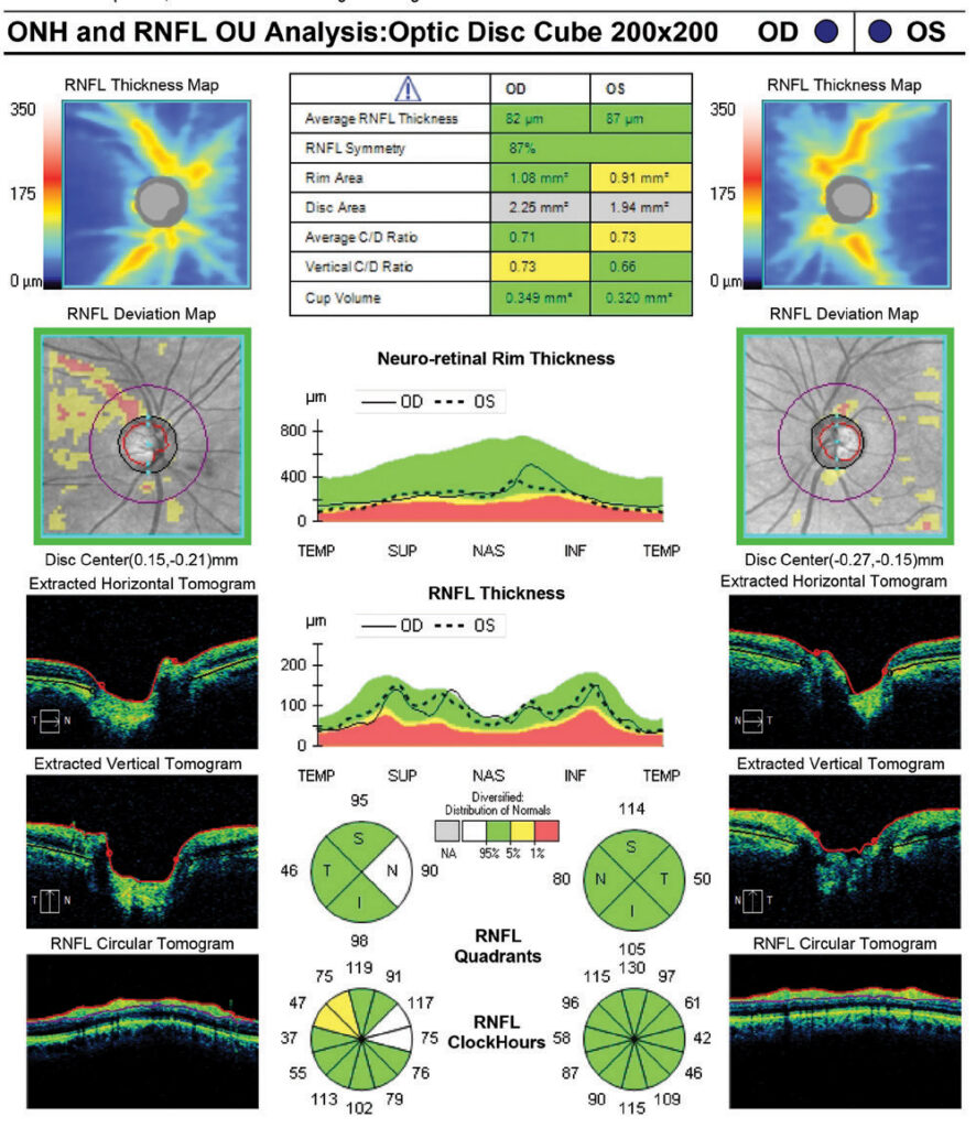
Additional image: Optical coherence tomography (OCT)

01

Optical coherence tomography (OCT)

Purpose: to obtain cross-sectional retinal images and quantitative measurements of retinal and optic nerve structures without touching the eye.

How it works and why it is used

Spectral-domain OCT uses low-coherence light interferometry to generate retinal cross-sections. Depending on the scan protocol, retinal layers and optic nerve structures can be measured and compared over time. Resolution and measurements depend on the platform and protocol.

Clinical applications

Macular abnormalities, oedema, epiretinal membranes, macular holes, age-related macular degeneration, diabetic retinal changes and retinal nerve fibre layer loss in glaucoma or other optic neuropathies.

During the examination

You rest your chin and forehead on the supports and look at a fixation target for a few seconds. Scanning is non-invasive, contact-free and painless.
Ocular biometry at Axa Optic
Eye examination equipment at Axa Optic
Additional example: Ocular biometry
Additional image: Ocular biometry

02

Ocular biometry

Purpose: to measure axial length and other ocular parameters used to calculate intraocular lens power and assess the eye’s anatomy.

How it works and why it is used

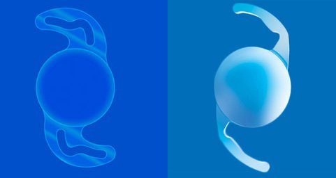
Optical biometry measures axial length and, depending on the instrument, anterior chamber depth, lens thickness and related parameters. Together with keratometry and an appropriate formula, measurements help estimate the intraocular lens power before cataract surgery.

Clinical applications

Axial myopia, differences in eye length, anterior segment measurements and parameters relevant to cataract surgery planning and intraocular lens selection.

During the examination

You look at a fixation target for a few seconds without contact with the eye. If ultrasound biometry is required, the clinician will explain the technique.

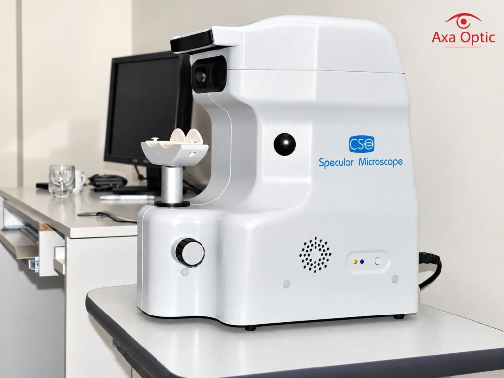
Axa Optic — Corneal endothelial specular microscopy
Corneal endothelial specular microscopy
Additional example: Corneal endothelial specular microscopy
Additional image: Corneal endothelial specular microscopy

03

Corneal endothelial specular microscopy

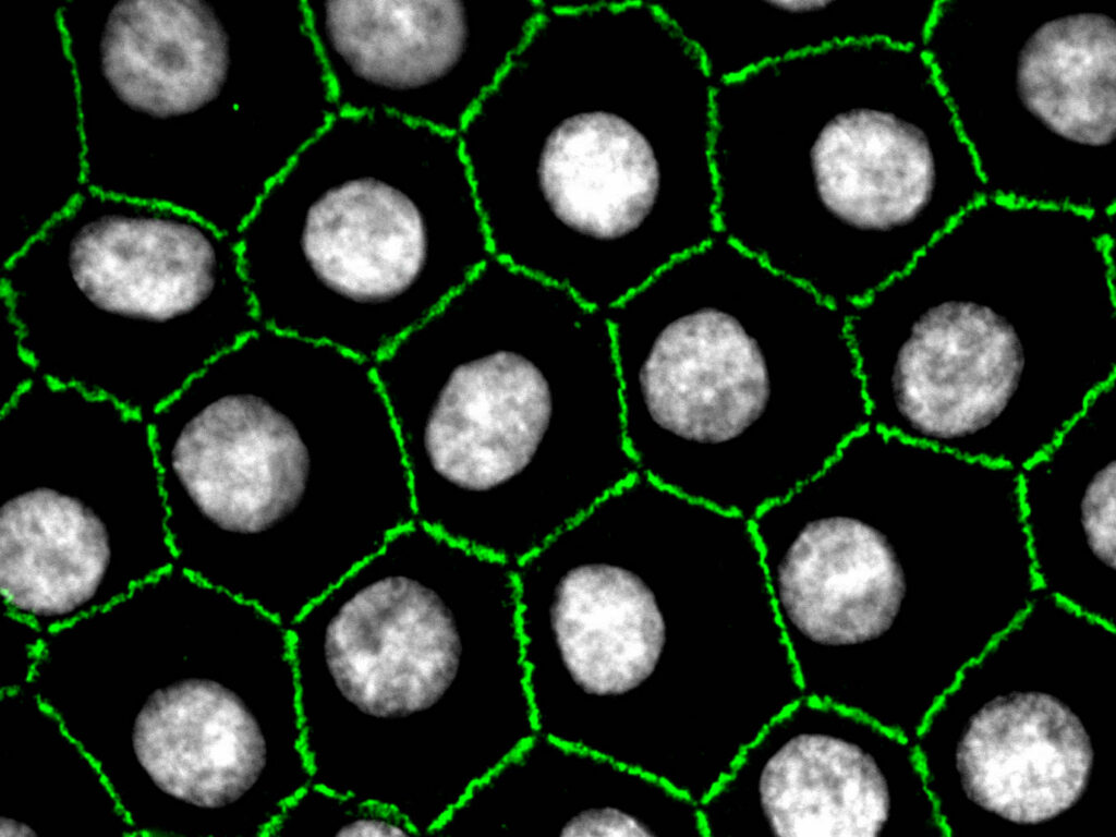
Purpose: to image and assess the corneal endothelium, the cell layer responsible for maintaining corneal transparency.

How it works and why it is used

Specular microscopy assesses endothelial cell density and morphology, including variation in cell size and the proportion of hexagonal cells. Endothelial reserve is important when evaluating risks associated with certain intraocular procedures.

Clinical applications

Endothelial cell loss or morphological changes, suspected Fuchs endothelial dystrophy, and assessment before or after indicated intraocular surgery.

During the examination

You look at a fixation target while the device takes microscopic images of your cornea. The test is normally quick, non-contact and painless.

Axa Optic — Ocular ultrasonography (B-scan)
Ocular ultrasonography (B-scan)
Additional example: Ocular ultrasonography (B-scan)
Additional image: Ocular ultrasonography (B-scan)

04

Ocular ultrasonography (B-scan)

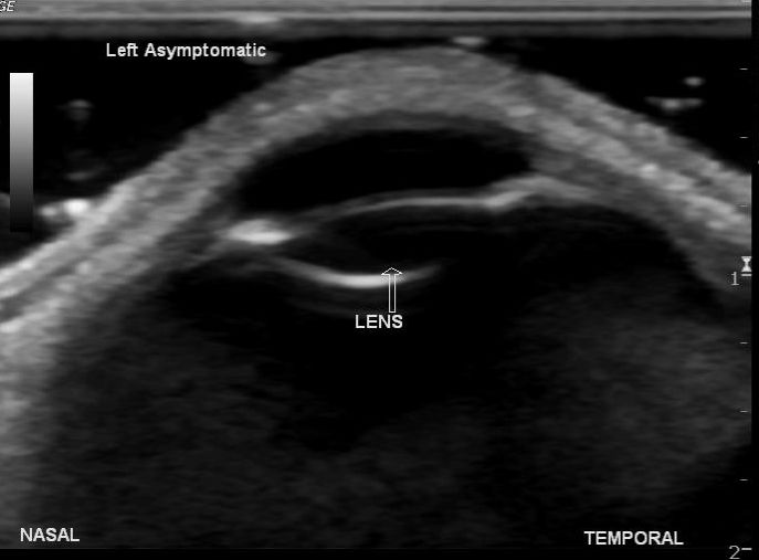
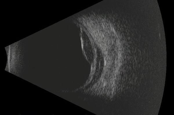
Purpose: to assess structures inside and behind the eye using ultrasound, particularly when direct examination is limited.

How it works and why it is used

B-scan ultrasound provides complementary information about the globe and posterior segment, including when cataract or other media opacities prevent direct fundus examination. It answers different clinical questions from OCT.

Clinical applications

Retinal detachment, vitreous haemorrhage, vitreous opacities, certain intraocular masses and other posterior segment abnormalities.

During the examination

Depending on the technique, scanning is performed through the eyelid or with ocular contact after topical anaesthetic. A suitable coupling medium is used.

Axa Optic — Automated visual field testing
Automated visual field testing
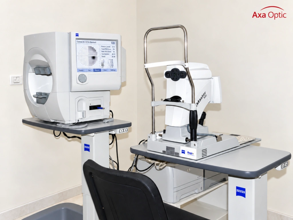
Visual field and glaucoma investigation at Axa Optic
Visual field testing and glaucoma assessment

05

Automated visual field testing

Purpose: to assess sensitivity across the central and peripheral visual field.

How it works and why it is used

Automated perimetry presents controlled light stimuli at predefined locations. Sensitivity values, global indices and deviation maps help assess functional loss and changes over time.

Clinical applications

Visual field defects related to glaucoma, some retinal conditions, optic nerve disorders and selected neurological disorders.

During the examination

You keep looking at a central target and indicate when you see a light. The test requires attention and cooperation.

Axa Optic — Fundus photography
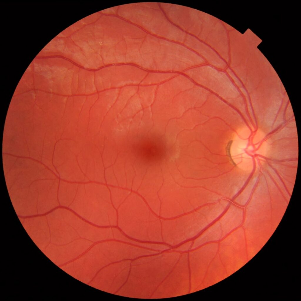
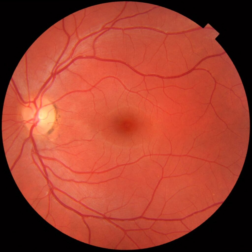
Fundus photography
Additional example: Fundus photography
Additional image: Fundus photography

06

Fundus photography

Purpose: to document the retina, macula, retinal vessels and optic nerve head with detailed photographs.

How it works and why it is used

The field of view and image resolution depend on the camera and acquisition protocol. Photographs allow the clinician to document retinal findings and compare them over time.

Clinical applications

Diabetic retinopathy, vascular changes, macular lesions, age-related macular degeneration, optic nerve changes suggestive of glaucoma and other retinal abnormalities.

During the examination

You look at the camera target while a brief light exposure records the image. Pupil dilation may be advised for some examinations.

Non-contact tonometry at Axa Optic
Eye examination equipment at Axa Optic

07

Non-contact tonometry

Purpose: to estimate intraocular pressure without touching the cornea.

How it works and why it is used

Intraocular pressure is expressed in mmHg. Corneal thickness and biomechanics can influence readings, especially with non-contact methods; a pressure reading alone cannot establish a glaucoma diagnosis.

Clinical applications

Detection and follow-up of increased or reduced intraocular pressure and identification of findings that require a further ophthalmological assessment.

During the examination

You fixate on a target while a short puff of air is directed towards the cornea. The measurement takes only a few seconds.

Autokeratorefractometry at Axa Optic
Eye examination equipment at Axa Optic

08

Autokeratorefractometry

Purpose: to provide initial objective estimates of refraction and corneal curvature.

How it works and why it is used

Autorefraction gives an initial objective measurement of refractive error, while keratometry measures the curvature of the principal corneal meridians. Results must be correlated with clinical refraction.

Clinical applications

Myopia, hyperopia, astigmatism, contact lens assessment and some measurements used in preoperative calculations.

During the examination

You look at the device target while the measurement is taken automatically, without touching the eye.

Interpreting your results

Investigations provide objective measurements and images. Your physician establishes the diagnosis by considering the results alongside symptoms, medical history and a clinical eye examination.

Request an appointment
Scroll to Top