 ®
Blurred vision
The cloudy lens no longer transmits and focuses light as effectively, reducing the clarity of the image formed on the retina.
Cataract
A cataract is the progressive loss of transparency of the eye’s natural lens. As it becomes cloudy, light is transmitted and focused less effectively on the retina, potentially reducing visual clarity, contrast and quality of vision. [1][2]
Cataract removal is surgical: the cloudy natural lens is removed and replaced with an intraocular lens (IOL). The indication, timing and choice of implant are individualized after an ophthalmic examination. [1]
01 · Early signs · consultation and diagnosis
The natural crystalline lens is a transparent structure behind the iris that helps focus light on the retina. Cataracts develop when the lens progressively loses transparency. Light is then transmitted and focused less effectively. [1][2]
The course is usually gradual and may differ between the eyes. Changes in the lens can cause blurred vision, halos, glare, reduced contrast sensitivity and changes in refractive error. [1][2]
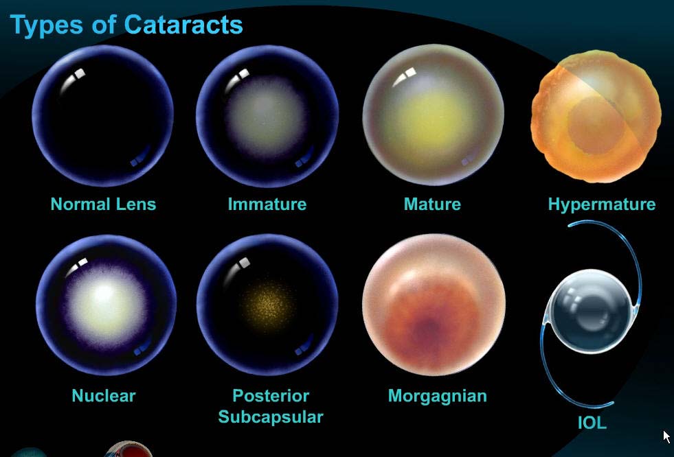
You do not need to wait until vision becomes severely impaired. An examination is appropriate when visual changes begin to affect daily activities, work or driving safety. [1][2]
Symptoms and optical mechanisms
The cloudy lens no longer transmits and focuses light as effectively, reducing the clarity of the image formed on the retina.
Cloudy areas of the lens scatter light, so headlights, lamps and other bright sources may become more bothersome, particularly in low-light conditions.
Changes in light transmission through the lens can make subtle differences between shades and colors harder to distinguish.
As the optical properties of the lens change, so can the eye’s refraction. Spectacle prescriptions may therefore change more frequently before cataracts become obvious to the patient.
An uncomplicated cataract usually causes gradual rather than sudden vision loss. Sudden loss of vision, significant eye pain or marked redness should not automatically be attributed to cataracts and require prompt ophthalmic assessment.
The consultation
The ophthalmic consultation assesses lens opacity, visual function and any other conditions that could affect vision or the outcome of surgery. [1]
Diagnosis and indication for surgery
The ophthalmologist confirms the diagnosis by examining the lens with a slit lamp and relating its appearance to visual function. [1]
Surgery is considered when cataracts explain visual impairment that sufficiently affects the patient’s activities for the expected benefit to outweigh the risks and inconvenience of the procedure. [1]
The decision to operate is not based on a single visual-acuity threshold. The decision takes account of symptoms, cataract severity, visual function, patient needs, ocular and systemic comorbidities, and the realistically achievable visual outcome. [1]
02 · Investigations, laboratory tests and medical clearance
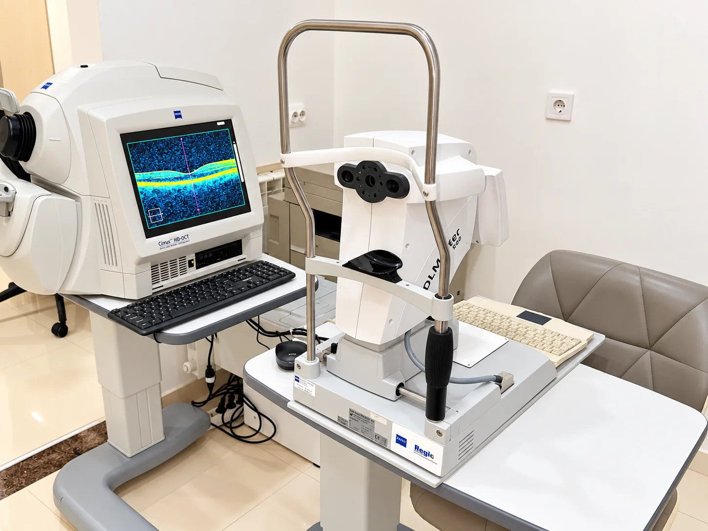
Before surgery, the ocular measurements needed to calculate and select the IOL are assessed separately from general health and perioperative risk. Ophthalmic investigations and medical assessment serve different, complementary purposes. [1][3]
Measurements and imaging for IOL power calculation, corneal assessment, detection of retinal disease or other factors limiting outcomes, and selection of the refractive target.
Medical history, current medication, laboratory tests, ECG, specialist clearance and pre-anesthetic consultation according to the clinical protocol and the patient’s circumstances.
Core measurements
Optical biometry measures axial length and, depending on the device, parameters such as anterior chamber depth, lens thickness and corneal diameter. These measurements are used in modern formulas for calculating intraocular lens power. [1][10]
Situations requiring additional care include very short or long eyes, previous corneal refractive surgery and inconsistent measurements.Keratometry measures corneal power and astigmatism and is essential for IOL calculation and selection of a toric strategy when astigmatism is relevant. [1][9]
Measurements must be reproducible; unstable or inconsistent values should be rechecked before the final decision.Preoperative refraction describes the current optical state but does not by itself determine IOL power. The postoperative refractive target is a separate decision, discussed in relation to eye anatomy, implant type, activities and tolerance for spectacles. [1]
IOL dioptric power and the postoperative refractive target are different parameters.Measurement quality
Tear-film instability, dry eye and other corneal surface irregularities can alter keratometry and consequently IOL calculation. Significant ocular surface disease should be treated and final measurements repeated after stabilization. [8]
Additional investigations
Useful when astigmatism requires more precise characterization, before a toric IOL, in irregular corneas or after refractive surgery. Assessment of the posterior corneal surface can improve estimation of total astigmatism. [9][10]
OCT may reveal epiretinal membranes, macular degeneration, vitreomacular traction, edema or other lesions not always apparent on fundus examination. These findings can affect the visual prognosis and choice of IOL. [7]
It is particularly relevant when vision is worse than the cataract would explain or when a presbyopia-correcting implant is being considered.Indicated when glaucoma is suspected or diagnosed, the optic disc appears abnormal, or other findings may limit functional outcomes. Results are interpreted in the context of the clinical examination and intraocular pressure. [1]
Measures corneal endothelial cell density and morphology. Important in the presence of guttae, endothelial dystrophy, corneal edema, previous eye surgery or other signs of reduced endothelial reserve. [1]
In very dense cataracts, when the retina cannot be examined adequately, B-scan ultrasonography can assess the posterior segment. A-scan ultrasound may be used for biometry when optical biometry is not feasible or sufficiently reliable. [1]
After LASIK/PRK or in eyes with extreme anatomical dimensions, IOL calculation requires specific formulas and strategies. Previous refractive surgery must be disclosed even if it took place many years earlier. [10]
When a toric or presbyopia-correcting implant is being considered
For a toric IOL, the magnitude and axis of astigmatism, corneal regularity, posterior corneal contribution and implant alignment are important. For EDOF or multifocal/trifocal IOLs, the macula, optic nerve, cornea, ocular surface and patient expectations must also be assessed, because coexisting eye disease may reduce the benefit of a more complex optical design. [1][7][9]
Axa Optic protocol
This list reflects the Axa Optic clinical protocol and should be distinguished from general recommendations in international guidelines. Additional tests or specialist opinions may be requested based on medical history and the pre-anesthetic assessment.
For cataract surgery, available evidence does not show that routine universal medical testing for every patient reduces perioperative complications. Guidelines and research support assessment tailored to the patient and clinical context, while each facility may follow its own operational protocol. [6]
At Axa Optic, the preoperative checklist standardizes preparation and ensures that the surgical and anesthesia teams have the necessary information before the procedure.
Information to share with the medical team
Anticoagulants, antiplatelet drugs, medication for diabetes, hypertension or prostate conditions, and all other long-term treatments must be disclosed. Do not stop medication on your own.
LASIK/PRK, injuries, glaucoma, uveitis, intravitreal injections, retinal surgery and other procedures may affect investigations and surgical planning.
Cardiovascular, respiratory, neurological and kidney conditions, diabetes, medication allergies and previous reactions to anesthesia should be reported in full.
Tremor, severe anxiety, difficulty lying flat, breathlessness, neck or back pain and communication difficulties may influence anesthesia choice and surgical arrangements.
Do not stop or change treatment before surgery without explicit medical advice. For anticoagulants, antiplatelet drugs, diabetes medication and other treatments affecting perioperative care, the clinicians involved determine the appropriate plan individually.
03 · Intraocular lens implantation
After the cloudy natural lens is removed, an intraocular lens (IOL) provides an essential part of the eye’s optical power. IOL selection involves calculating lens power, choosing the refractive target and optical design, and correcting astigmatism where appropriate. The decision takes account of ocular anatomy and the patient’s visual needs. [1][4][5]
The condition of the cornea, macula, optic nerve, pupil and ocular surface, together with astigmatism and other comorbidities, affects IOL selection and visual prognosis.
The main goal may be distance vision, monovision or mini-monovision, or a wider range of focus.
Night driving, computer work, reading, sports, professional requirements and tolerance for spectacles are relevant to IOL selection.
Depending on the optical design, reduced dependence on spectacles may come with halos, other light-related visual phenomena or reduced contrast sensitivity. These effects should be discussed before choosing an IOL. [4]
Optical lens families
Provides one main focal point. The most common goal is clear distance vision, with reading glasses usually needed for near tasks. Monofocal IOLs can also be used for monovision or another refractive target if discussed and accepted before surgery.
Provides a straightforward optical profile and good performance at the selected refractive target. When both eyes are targeted for distance, reading glasses are usually needed.A predominantly monofocal design optimized to extend performance, particularly at intermediate distance. Meta-analyses show a modest but significant improvement in intermediate vision compared with conventional monofocal IOLs, without making this a true multifocal design. [5][13]
May improve intermediate vision compared with conventional monofocal IOLs; near vision remains variable and glasses may still be needed.Extended depth of focus (EDOF) designs aim to provide a more continuous range of focus, mainly from distance to intermediate vision. On average, EDOF lenses offer better intermediate vision than monofocal lenses, while unaided near vision is generally weaker than with trifocal lenses. [4][13]
Extends depth of focus from distance toward intermediate vision; fine near tasks may still require glasses.Distributes light energy among multiple focal points to support distance, intermediate and near vision. Comparative studies report greater spectacle independence and better near vision than monofocal IOLs, but also more halos and other dysphotopsias. [4]
May increase spectacle independence at several distances, with a greater likelihood of halos and other dysphotopsias than monofocal IOLs.Toric IOL: correction of corneal astigmatism
A toric implant has different optical powers in different meridians to correct corneal astigmatism. Depending on the model, toricity may be available in monofocal, enhanced monofocal, EDOF or trifocal IOLs. Important factors include astigmatism magnitude and axis, posterior corneal contribution, IOL calculation and rotational stability. [9][11][12]

Illustrative clinical comparison

| IOL family | Distance | Intermediate | Near | Spectacle independence | Light-related visual phenomena |
|---|---|---|---|---|---|
| Monofocal | Very good at the selected target | Limited / variable | Usually limited | low | usually low |
| Enhanced monofocal | Very good at the selected target | Better than conventional monofocal | Limited / variable | low to moderate | generally similar to monofocal |
| EDOF | Good | Extended | Variable; fine near tasks may require glasses | moderate | varies by technology |
| Trifocal | Distance focal point | Intermediate focal point | Near focal point | higher | more common than with monofocal IOLs |
The table describes general trends across optical families. Outcomes depend on the specific IOL model, biometry, refractive target, ocular disease, binocular vision and individual adaptation. [4][5][13]
Patient selection
Presbyopia-correcting IOLs are not automatically ruled out by every comorbidity, but macular disease, optic neuropathy, significant glaucoma, irregular corneas, unstable ocular surfaces and other causes of reduced contrast require caution and a realistic discussion of expected benefits. [1][7][8]
Refractive target
A common strategy with monofocal IOLs. It maximizes symmetry for distance vision but usually requires optical correction for near tasks.
One eye is targeted closer to emmetropia and the other left slightly myopic to extend functional vision. This requires binocular compatibility and acceptance of the trade-off between eyes.
EDOF and trifocal lenses extend the range of focus through their optical design. This does not remove the need for accurate refractive calculations and control of residual astigmatism.
Axa Optic® Vision Explorer
Vision Explorer illustrates the principles of different optical strategies. The images do not reproduce an individual patient’s exact vision or simulate any specific commercial implant.
Usually prioritizes distance vision; reading glasses are often needed.



Realistic expectations
Even with accurate biometry and uncomplicated surgery, residual refractive error, residual astigmatism, glasses for certain activities or light-related visual phenomena may occur. Results also depend on the retina, optic nerve, cornea, ocular surface and binocular visual processing. [1][4]
The specific implant model, IOL power, toricity and refractive target are selected only after integrating preoperative measurements, clinical examination and the patient’s visual goals. The simulator and tables on this page are educational tools, not prescriptions.
04 · Preparing for surgery
Once the indication for surgery and implant plan have been established, general health, current treatments, requested tests and medical clearances are reviewed. Any change in health between the consultation and surgery must be reported to the medical team. [1]
In the days beforehand
On the day of surgery
On arrival at the clinic
Long-term medication
Cataract surgery generally carries a low bleeding risk. Perioperative management of anticoagulants and antiplatelet drugs depends on the reason for treatment, the medicine, INR where relevant, and the anesthesia technique. Unnecessary interruption can increase thromboembolic risk. [1][14]
Do not stop aspirin, clopidogrel, warfarin or direct oral anticoagulants without explicit medical advice.Insulin and diabetes medication schedules may need adjustment according to surgery time, food intake and the anesthesia plan. Tell the team exactly what you take and follow your individual instructions.
Do not change doses yourself on the morning of surgery.Tamsulosin and other alpha-blockers are associated with intraoperative floppy iris syndrome (IFIS), which can make surgery more difficult. Risk may persist even if the medicine was stopped previously, so disclosing its use is more important than stopping treatment without advice. [16]
Tell the doctor if you currently take or previously took tamsulosin or other medication for urinary or prostate symptoms.List antihypertensives, cardiac, respiratory and neurological treatments and all other long-term medicines. For some drugs, the time of the last dose and whether treatment has continued are important.
Bring an up-to-date list of medicine names, doses and dosing frequency.Food and fluids
In cataract surgery under topical anesthesia without sedation, a randomized trial reported comparable perioperative outcomes without fasting, with less anxiety and pain. This finding does not replace the anesthesia protocol; individual food and fluid instructions take priority. [15]
Eye drops and preoperative treatment
Preoperative medication is prescribed individually or according to the applicable clinical protocol. Medicines, concentrations, frequency and timing may differ by case; follow the written schedule provided to you.
Apply drops to the correct eye, at the prescribed times and doses. Do not add medicines preventively on your own.
Do not automatically double a missed dose. Continue according to your instructions or clarify the schedule with the medical team.
Unusual symptoms, allergic reactions, significant pain or discharge after medication should prompt contact with the clinic before changing treatment.
Changes to report to the clinic before surgery
Checklist for surgery day
General information on this website does not replace individual preoperative advice. If it differs from the written instructions of your surgeon or anesthetist, follow the individual instructions and clarify any uncertainty with the clinic.
05 · Surgery
At Axa Optic® Clinic, 185 Republicii Street, Bârlad, the standard cataract procedure is phacoemulsification with implantation of an artificial intraocular lens. Through a small incision, the cloudy lens is broken up and aspirated, and a foldable IOL is generally placed in the capsular bag. The final plan may be adapted during surgery if ocular anatomy or patient safety requires it. [1]
Immediate preoperative checks
The team verifies patient identity, the eye to be operated on, planned IOL type and power, allergies, relevant medicines and anesthesia. Any discrepancy must be resolved before the procedure starts.
Asepsis and monitoring
The eye and surrounding skin are prepared with antiseptic, sterile drapes are applied, and the patient is monitored according to medical protocol. Infection prophylaxis follows the clinical protocol and applicable guidelines. [18]
Anesthesia
Given as eye drops, with the option of adding intracameral anesthetic if the surgeon considers it necessary. The patient remains awake and may perceive light, colors, movement or pressure; these sensations do not mean that they are watching the operation.
Topical anesthesia avoids the risks of a periocular injection but requires good cooperation and may need additional pain relief. [17]Anesthetic is injected around the eyeball to provide deeper pain relief and reduce eye movement. It may be preferred when cooperation is difficult, surgery is expected to be more complex or the doctor considers it provides better control.
It may cause bruising, swelling or bleeding around the eye and, rarely, more serious injection-related complications; its use is therefore individualized. [17]Possible sensations during surgery
During surgery, patients may perceive bright light, colors, water or pressure sensations and sounds from the equipment. If pain, marked anxiety, a need to cough or a need to change position occurs, tell the team and avoid sudden head or eye movements.
If pain relief is inadequate or cooperation becomes difficult, anesthesia may be supplemented or the technique changed. In unusual situations, surgery may be postponed or the patient referred to a center with additional support if that is safer.
Surgery step by step
One or more small corneal or limbal incisions are made to introduce instruments. A viscoelastic substance protects intraocular structures and maintains the working space.
The surgeon makes a controlled circular opening in the anterior lens capsule. Its shape and position matter for access to the cataract and subsequent IOL stability.
Depending on anatomy and technique, lens material is carefully separated from the capsule to allow safe fragmentation and removal.
The cloudy lens nucleus is fragmented and emulsified using ultrasound energy, then aspirated. Equipment settings and fragmentation technique are adapted to cataract density, anterior chamber depth and capsular-zonular stability. [1]
Residual cortex is removed by irrigation and aspiration; the capsular bag is inspected and prepared for the implant. The goal is to preserve a stable, clean and centered capsule where anatomy permits.
Viscoelastic material is aspirated and the surgeon checks IOL position, capsular bag stability, the anterior chamber and the integrity of intraocular structures.
Small incisions are usually self-sealing and may be hydrated to ensure closure; a suture may be used if necessary. Local treatment is administered according to protocol and the indicated eye protection is applied.
Intraoperative situations that may change the technique
Some situations, such as a small pupil, pseudoexfoliation, weak zonules, very dense cataract, previous eye surgery or IFIS, increase technical difficulty. Recognizing them beforehand allows contingency planning, but some become apparent only during surgery. [16][19]
Intracameral medicines, iris hooks or a mechanical pupil-expansion device may be needed to maintain a safe surgical field.
Capsular support devices and adapted techniques may be used to preserve bag stability and reduce traction on the zonules.
This may require anterior vitrectomy, a change in how lens material is removed and a different IOL position or type; implantation may sometimes be deferred.
If patient safety requires it, the surgeon may change the technique, stop the procedure or recommend further treatment at a center with additional resources.
Preventing infection
Periocular and ocular antisepsis, sterile draping and antibiotic prophylaxis according to protocol are central measures for reducing endophthalmitis risk. Randomized studies and systematic reviews support perioperative prophylaxis in cataract surgery. [18]
The substance, route and timing of antibiotic prophylaxis are determined by the surgical protocol, allergy history and case-specific factors.
Risks of surgery
Cataract surgery generally has a good safety profile, but no intraocular procedure is risk-free. Individual risk depends on ocular anatomy, cataract density, pupil dilation, zonular stability, eye comorbidities and previous surgery. Severe complications are uncommon but can permanently affect vision. [1][3]
The posterior capsule can tear during lens removal. Vitreous prolapse may occur, requiring anterior vitrectomy, a change in technique or a different IOL position or type. [1]
Zonular dialysis or weakness may destabilize the capsular bag. Nuclear fragments can enter the vitreous cavity and sometimes require additional vitreoretinal surgery. [1]
A small pupil, IFIS, iris prolapse or injury may require intracameral medicines, iris hooks or pupil-expansion devices and may prolong surgery. [16]
The corneal endothelium can be affected by ultrasound energy and intraocular maneuvers. Consequences may include prolonged corneal edema and, rarely, corneal decompensation. [1]
Significant intraocular bleeding is rare. Suprachoroidal hemorrhage is a severe complication that may require stopping surgery and additional surgical treatment. [1]
If the capsular bag cannot provide adequate support, the planned IOL may not be implanted in the intended position. Another implant type, alternative fixation or, rarely, delayed implantation may be required. [1]
At the end of surgery
At completion, eye stability, implant position and incision sealing are checked. Vision may be temporarily blurred by pupil dilation, medication, mild edema or an eye shield. The result is assessed progressively during follow-up visits.
The patient receives postoperative instructions, a treatment schedule and follow-up appointments. Significant pain, marked redness or sudden vision loss are not part of the expected recovery and must be reported promptly.
The surgical technique, anesthesia and IOL position may be changed during surgery if ocular anatomy or a complication requires it.
06 · Postoperative treatment
After cataract surgery, patients receive a written treatment schedule and instructions tailored to the operated eye, procedure and risk profile. Individual postoperative instructions take priority. [1]
Some postoperative inflammation is expected. Topical anti-inflammatory treatment aims to reduce inflammation and the risk of complications such as cystoid macular edema. Depending on the case, treatment may include a topical corticosteroid, a nonsteroidal anti-inflammatory drug (NSAID), or both. [20]
The incisions are small, but the eye is healing. Hand hygiene and avoiding eye rubbing, contaminated water, dust, smoke and direct contact with cosmetics reduce unnecessary exposure to irritants and contaminants.
Eye protection, physical activity and resuming driving are decided individually. Axa Optic recommendations include avoiding pressure on the eye, strenuous exercise, sudden bending, heavy lifting, swimming pools and saunas until cleared by the doctor.
Eye drop schedule
Frequently used to control intraocular inflammation after surgery. Dosing frequency and tapering depend on clinical examination; do not extend, stop or restart a corticosteroid on your own. [1][20]
May be prescribed alone or with a corticosteroid, especially to reduce the risk of pseudophakic macular edema. Meta-analyses show lower rates of macular edema with regimens including NSAIDs, but the optimal protocol varies between patients. [20]
If included in the prescribed regimen, use the antibiotic at the indicated dose, frequency and duration. Do not reuse medication left over from a previous procedure without medical advice.
Dryness, foreign-body sensation and tear-film instability may occur or temporarily worsen after surgery. If necessary, the doctor may add ocular surface treatment; no single regimen suits every patient. [21]
How to use eye drops
Follow the prescribed schedule; clarify any uncertainty about continuing treatment with the clinic.
Do not change subsequent doses on your own; follow the advice given for the situation.
The first few days
Watery eyes, mild foreign-body sensation, slight discomfort, some remaining blurred vision, light sensitivity and moderate redness may occur after surgery and should be considered in the context of overall recovery.
May be influenced by pupil dilation, mild corneal edema, unstable tear film or early postoperative inflammation.
May arise from the ocular surface, incision or dryness and is usually temporary. [21]
May occur temporarily, particularly during the first few days.
Some conjunctival redness may persist initially and should be assessed at scheduled follow-up visits.
Possible postoperative complications
Most patients recover without severe complications. Nevertheless, cataract surgery can be followed by inflammatory, infectious, corneal, retinal, refractive or IOL-related complications. Some require rapid treatment to protect vision. [1][3]
A rare but severe intraocular infection that can cause pain, redness, discharge and rapid vision loss. It requires emergency ophthalmic assessment and treatment. [24]
Postoperative inflammation can be more intense than expected. Toxic anterior segment syndrome (TASS) is a rare sterile inflammatory reaction that must be rapidly distinguished from endophthalmitis. [1]
Intraocular pressure may rise temporarily or persistently after surgery. Risk and management depend on measured pressure, glaucoma and other ocular factors. [1]
Early corneal edema can delay visual recovery. In patients with reduced endothelial reserve, it may persist and rarely progress to pseudophakic bullous keratopathy. [1]
May reduce or distort vision several weeks after surgery. OCT often confirms the diagnosis, and treatment is tailored to severity and risk factors. [20]
A rare but important complication. Sudden onset of numerous floaters, flashes or a shadow or curtain across vision requires urgent retinal assessment. [1]
Decentration, tilt, rotation of a toric IOL, dislocation or late displacement can occur. Implant repositioning or replacement is sometimes necessary. [1]
After stabilization, residual myopia, hyperopia or astigmatism may remain. Halos, glare and other dysphotopsias can persist, particularly with certain IOL designs. [1]
May develop months or years after surgery and reduce visual acuity and contrast again. When visually significant, it may be treated by Nd:YAG laser capsulotomy. [22][3]
Capsular fibrosis and contraction can alter implant position. Very rarely, the IOL material may become cloudy or calcify, requiring surgical assessment. [1]
sudden or marked vision loss, significant or increasing pain, severe redness, discharge, intense light sensitivity, sudden onset of many floaters, flashes or a curtain/shadow across the visual field. See warning signs and recommended action.
Protecting the eye and hygiene
Postoperative recommendations include avoiding rubbing or direct pressure on the operated eye.
Wash carefully, minimizing direct contact of the eye with contaminated water, shampoo, soap or cosmetics until normal routines are permitted by the doctor.
Use an eye shield or other protection during sleep if recommended in your postoperative instructions.
Axa Optic postoperative advice includes temporarily avoiding dusty or smoky environments, swimming pools and saunas until cleared by the doctor.
Physical activity and daily life
Light activities are generally compatible with early recovery unless the doctor advises otherwise. The timing of strenuous exercise, heavy lifting, repeated bending, sports, swimming and activities that risk eye injury is determined at follow-up.
May be resumed early if general health and vision allow.
Resume driving only after medical confirmation that your vision and eye condition make it safe.
Resume after medical clearance, depending on the activity and postoperative course.
May be used as comfort permits; they do not mechanically affect the implant. Breaks may help if fatigue or dry eyes occur.
Ocular surface after cataract surgery
Dry-eye symptoms and tear-film instability may persist for weeks or months after cataract surgery. Their persistence does not itself indicate an IOL problem; the ocular surface should be assessed separately and treatment tailored to findings. [21]
Lubricants or other ocular surface therapies may be helpful in some cases, but antibiotics, corticosteroids and other active medicines are not automatically added to the postoperative regimen.
Treatment may change at follow-up
Follow-up examinations assess inflammation, the cornea, intraocular pressure, macula and visual recovery. Based on these findings, the doctor may reduce, extend, change or supplement treatment. The postoperative regimen and follow-up visits are part of the same care plan. [1]
Axa Optic postoperative instructions specify the operated eye, surgery date, prescribed treatment, next appointment and contact details. The instructions given to the individual patient take precedence over general examples or regimens used by others.
Doses, frequency and duration of postoperative treatment should change only according to clinical progress and medical advice.
07 · Follow-up visits and warning signs
After cataract surgery, recovery should be monitored until the eye and refractive result have stabilized. Follow-up helps detect excessive inflammation, intraocular pressure changes, corneal or macular edema, implant-position problems and other complications that may require a treatment change. [1][20]
The doctor checks corneal clarity, inflammation, intraocular pressure, implant position and changes in visual acuity.
Eye drops may be reduced, extended, changed or supplemented based on examination findings and individual risk factors.
The final optical prescription is determined once refraction and the ocular surface are sufficiently stable for reliable measurements.
Follow-up schedule
The schedule depends on surgical complexity, anesthesia, comorbidities and postoperative progress. The date, time and location are recorded in the patient’s postoperative documents.
Assesses the cornea, inflammation, intraocular pressure, implant position and any changes requiring treatment adjustment.
Monitors healing, response to treatment, ocular surface, functional vision and possible signs of edema or persistent inflammation.
Assesses the postoperative result, refraction, need for spectacles and whether further investigations or treatment are required.
Arranged when recovery differs from expectations, risk factors are present or a particular ocular structure requires closer monitoring.
What is assessed at postoperative follow-up
Visual progress is monitored, distinguishing normal refractive recovery from other causes of vision below expectations.
May change after surgery and require treatment or additional monitoring, particularly in patients with glaucoma or other risk factors.
The doctor checks transparency, edema, incision integrity and the ocular surface. Corneal recovery directly affects visual clarity in the first days and weeks.
Inflammation in the anterior chamber and response to prescribed treatment are assessed.
IOL centration, stability and position are checked; in toric implants, axis orientation can be important for the refractive result. [11][12]
If vision is worse than anterior-segment findings explain, or specific symptoms arise, retinal examination and OCT may identify macular edema or other causes of reduced vision. [20]
Warning signs
Significant eye pain, sudden or marked vision loss, severe redness, discharge, intense light sensitivity, nausea or vomiting associated with eye pain, and injury to the operated eye require prompt contact with the clinic or urgent ophthalmic assessment. [1]
sudden loss, rapidly worsening blur or a shadow/curtain across the visual field
severe or increasing pain, or pain with headache, nausea or vomiting
severe or progressive redness, substantial discharge, marked eyelid swelling or intense light sensitivity
sudden onset of many floaters, flashes of light or a curtain/shadow across the visual field
a blow to the operated eye or any event followed by changes in vision, pain or the appearance of the eye
Postoperative warning signs
Pain, redness and vision loss can occur with intraocular infection, severe inflammation, markedly raised intraocular pressure, corneal injury or other complications. Flashes, sudden onset of numerous floaters and a curtain-like shadow may indicate retinal disease. These symptoms require ophthalmic examination and cannot be reliably distinguished remotely.
Rescheduling a postoperative visit
Axa Optic postoperative instructions explicitly require patients to attend scheduled visits or notify the clinic in good time if they cannot attend. The reason is medical: some complications initially cause few symptoms but can be detected on examination before they significantly affect vision.
In the longer term
The natural lens has been removed and cannot develop another cataract. However, the posterior capsule supporting the implant may become cloudy over time, causing blurred vision, halos, glare and reduced contrast sensitivity. [22]
Visually significant posterior capsule opacification may be treated by Nd:YAG laser capsulotomy after an eye examination. The procedure creates an opening in the cloudy capsule to restore the visual axis, once it has been confirmed as the cause of reduced vision. [3]
After capsulotomy, flashes, a sudden increase in floaters or a curtain across the visual field require prompt retinal assessment. [23]
Visual recovery below expectations
May require spectacles, contact lenses or, selectively, other refractive options once the eye has stabilized.
May cause fluctuating vision and discomfort even when the implant is well positioned. [21]
Macular edema or pre-existing retinal disease can limit visual recovery and may require OCT or other investigations. [20]
Comorbidities may limit vision independently of the technical success of surgery and need separate monitoring.
Contacting the clinic promptly
Contact the clinic using the details in your postoperative documents. Severe or rapidly progressing symptoms, or inability to reach the clinic promptly, require emergency ophthalmic assessment.
Postoperative recovery is assessed through symptoms, visual acuity and clinical examination. Report any significant change between visits to the medical team.
This content is for information and does not replace a consultation, clinical examination or individual medical advice.
Cataract consultation
The consultation determines whether cataracts explain the symptoms, whether other causes of reduced vision are present and when surgery should be considered.